Saturday, September 8, 2007
Friday, September 7, 2007
Saturday, August 25, 2007
Epithelial Tissue
Occurring in sheets of tightly packed cells, epithelial covers the outside of the body and lines organs and cavities within the body. The cells of an epithelial tissue, or epithelium, are closely joined, with little material between them. In many epithelia, the cells are riveted together by tight junctions. This tight packing enables the epithelium to function as a barrier against mechanical injury, microbes, and fluid loss. Some epithelia, called glandular epithela, absorb or secrete chemical solutions. For example, the glandular epithelia that line the lumen of the digestive and respiratory tracts form a mucous membrane; they secrete mucus that lubricates the surface and keeps it moist.
Two criteria for classifying epithelia are the number of cell layers and the shape of the cells on the exposed surface. A simple epithelium has a single layer of cells, whereas a stratified epithelium has multiple tiers of cells. A "psuedostratified" epithelium is single-layered but appears stratified because the cells vary in length. The shape of the cells at the exposed surface may be cuboidal, columnar, or squamous.
Two criteria for classifying epithelia are the number of cell layers and the shape of the cells on the exposed surface. A simple epithelium has a single layer of cells, whereas a stratified epithelium has multiple tiers of cells. A "psuedostratified" epithelium is single-layered but appears stratified because the cells vary in length. The shape of the cells at the exposed surface may be cuboidal, columnar, or squamous.
Friday, August 24, 2007
Tissue Structure and Function
Tissues are groups of cells with a common structure and function. Different types of tissues have different structures that are suited to their functions. For example, a tissue may be held together by a sticky extracellular matrix that coats the cells or weaves them together in a fabric of fibers. In deed, the term tissue derives from a Latin word meaning 'weave.'
Tissues are classified into four main categories - epithelial tissue, connective tissue, muscle tissue, and nervous tissue.
Tissues are classified into four main categories - epithelial tissue, connective tissue, muscle tissue, and nervous tissue.
Friday, August 3, 2007
Light Microscopes

The microscopes first used by Renaissance scientists, as well as the microscopes you are likely to use in the laboratory, are all light microscopes (LMs). Visible light is passed through the specimen and then throught glass lenses. the lenses refract (bend) the light in such a way that the image of the specimen is magnified as it is projected into the eye, ont photographic film or a digital sensor, or onto a video screen.
Saturday, July 28, 2007
Supraspinatus Muscle

ACTION: abducts the humerus (lifts the arm) REFERRAL: lateral shoulder and arm
COMMENTS: This muscle runs under the clavicle, attaching to the top of the humerus. It pulls the arm out to the side. The supraspinatus and deltoid are the only two muscles that lift the arm to the side (the trapezius helps to stabilize and helps after 90 degrees). If strained, it can mimic subacromial bursitis. To work the muscle belly, you must work through the trapezius. This is one of the 4 rotator cuff muscles.
ROTATOR CUFF MUSCLES: (acronym SITS) Subscapularis, Infraspinatus, Teres minor, Supraspinatus
Thursday, July 26, 2007


Wednesday, July 25, 2007
Structure of Clavicle

- Sternal or medial end - the triangular-shaped, medial surface that articulates with the sternum. Use this marking as a landmark to identify the medial side of this bone.
- Acromial or lateral end - the flattened, lateral surface that articulates with the acromion of the scapula. Ise this marking as a landmark to identify the lateral side of this bone.
- Conoid tubercle - a small bump located on the inferior surface, near the acromial end, serves as an attachment point for a ligament. Ise this marking as a landmark to identify the inferior surface af this bone.
Tuesday, July 24, 2007


Monday, July 23, 2007

Friday, July 20, 2007
Serratus Anterior as the cause of scapula dysrhythmia
As Jon JP Warner et al (1998) explain: "The serratus anterior functions to maintain scapular stability during arm elevation. It does this by causing upward rotation and protraction of the scapula. Dysfunction of this muscle will cause winging of the scapula as the patient attempts to elevate the arm".
The long thoracic nerve is formed from the anterior primary rami of the 5th and 6th cervical vertebrae (C5 and C6). A third, or even a fourth branch, can originate from C4 and C7. It is purely a motor nerve and is the sole innervation to the serratus anterior muscle. The 5th and 6th cervical roots, along with the dorsal scapular nerve, pass through the substance of scalenus medius, a muscle in the side of your neck, whereas the seventh root passes anterior to it. The nerve passes downwards, beneath the brachial plexus and collar bone, over the first rib and on to the digitations of the serratus anterior along the outer side of the chest wall. Each digitation of the serratus anterior in innervated by an individual branch of the long thoracic nerve. the nerve can extend down to the 8th or 9th rib. Because the nerve is long and superficial it is very vulnerable to injury!
The serratus anterior has three functional components (Gregg et al 1979, Jobe 1998). The uppermost originates from the first two ribs and inserts on the superior angle of the scapula. The middle component originates from the 2nd, 3rd and 4th ribs, and inserts along the anterior aspect of the medial scapular border. The lowermost, and largest, originates from the 5th to 9th ribs. These digitations converge to insert on the inferior angle of the scapula.
During normal movement, particularly movements involving pushing, the scapula slides over the rib cage, and is held in place by the serratus anterior. If weakness or paralysis of the serratus is present, the scapula stands prominent from the rib cage when the arm is protracted against resistance. The muscle is usually tested by elevating both arms and pressing forwards against a wall (standing press-up). In synergy with the trapezius, the serratus anterior acts to provide a strong, mobile base of support to position the scapula optimally for maximum efficiency of the upper limb. As has already been stated, without the upward rotation and protraction of the scapula by serratus anterior, the arm can not be elevated fully. Gregg et al (1979) report that abduction is limited to110 degrees.
In the January/February 2000 edition of the Journal of Shoulder and Elbow Surgery, Hester et al describe a series of dissections undertaken to evaluate the course of the nerve. They make the following important observations: "In all specimens a tight fascial band of tissue arose from the inferior aspect of the brachial plexus, extended just superior to the the middle scalene muscle insertion on the first rib, and presented a digitation that extended to the proximal aspect of the serratus anterior muscle". As the next section reveals, this fascial band may be a cause of long thoracic nerve dysfunction!
Causes of winging scapula (scapula dysrhythmia)
1. Loss of serratus anterior muscle function
When one talks about winging of the scapula, true winging is due to serratus anterior muscle dysfunction. This is an uncommon condition and may arise from traumatic injury to the nerve supplying the serratus anterior muscle, the long thoracic nerve; or due to damage to the nerve from pressure lesions or a neuritis (inflammation of the nerve).The test for identifying a long thoracic nerve injury is the wall test. The patient is asked to face a wall, standing about two feet from the wall and then push against the wall with flat palms at waist level.
The treatment will depend on the cause and usually an MR scan is helpful to exlcude a mass lesion pressing on the nerve. See the excellent summary by Steven Fromm at http://freespace.virgin.net/steven.fromm/ for much more detail.
2. Loss of trapezius muscle function
The trapezius muscle is a large muscle above your scapula which lifts and rotates your scapula. It is the muscle you use to shrug your shoulders.Isolated loss of trapezius function is extremely rare and may occur after radical neck surgery (for tumours), where the nerve supplying trapezius may be damaged (the spinal accessory nerve).
3. Weakness of all the scapula stabilisers
Muscular dystrophies, most commonly fascioscapulohumeral dystrophy (FSHD), are the main cause of weakness of all the scapula stabilising muscles.
4. Loss of scapular suspensory mechanism
The coracoclavicular ligaments suspend the scapula from the clavicle and the acromioclavicular joint is the only joint linking the scapula to the rest of the body. Therefore dislocation of the acromioclavicular joint or a fracture of the outer third of the clavicle, with rupture of the coracoclavicular ligaments, leads to an abnormal scapula rhythm and apparent scapula winging with overhead manouevers. This is usually not painful and usually only affects overhead workers and athletes.
Another rare cause is the 'scapulothoracic dissociation', described by Rockwood & Matsen in 1990. The scapula is wrenched from the body in violent trauma leading to fracture of the clavicle and soft tissues around the clavicle.
5. Winging of the scapula secondary to instability
This is one of the commonest causes of scapula dysrythmia (and winging). Recurrent dislocations of the shoulder leads to dysfunction of the muscles that move and support the shoulder complex and scapula. The more frequent the dislocations and the less trauma involved in causing the dislocations, the worse the scapula dysryrhmia (winging). An essential part of treating shoulder instability (recurrent dislocations) is treating the scapula dysrythmia. This is done by an experienced physiotherapist in association with a shoulder surgeon.
6. Winging secondary to pain
This is another common cause of econdary winging and dysrhythmia of the scapula. Any painful condition of the shoulder will lead to abnormal movements of the entire shoulder complex. Reduced movement at the glenohumeral joint will lead to more compenatory movement at the scapula.
7. Brachial Plexus injury or disease
Most of the nerves supplying the stabilising muscles of the scapula arise from the Brachial Plexus. The Brachial Plexus is a bundle of nerves running from the neck to the arm. It carries the nerve supply for the muscles of the arm and shoulder. Sometimes a major accident can affect the muscles of the shoulder more than the arm and lead to winging. When there is no trauma, a condition known as Parsonage-Turner syndrome (Brachial Neuritis) can lead to weakness of the scapula muscles.
Wednesday, July 18, 2007
Breast Cancer Male
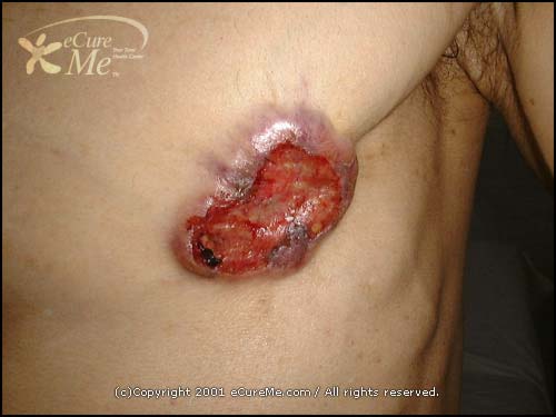
Only one case of Breast Cancer occurs in men for every 100 in women. The average age it occurs is 60. Male Breast Cancer is usually more aggressive (worse) than female Breast Cancer. Hormonal effects are thought to be the cause.
Symptoms:
Breast lump
Nipple discharge
Enlarged breast tissue
Nipple discharge
Enlarged breast tissue
Cause:
Probably hormonal
Liver damage may contribute since a damaged liver does not process hormones well, and there are higher levels of hormones in the blood. In some countries (Egypt, for example) where there is liver disease due to parasitic infections, the incidence of Breast Cancer in males is higher than in the United States.
There is a greater risk over the age of 65. It is rare under age 35.
Black men and men of European Jewish ancestry may have a higher risk than Caucasians.
How the diagnosis is made:
Examination
Hard breast mass beneath the nipple or areola (pigment area)
Enlarged lymph glands in the armpits, above collar bones, or in the center of the chest (mediastinal lymph nodes might be enlarged inside the chest, to the right and left of the midline)
Retracted/eroded nipple
Testing
Surgical biopsy
Bone scan to check for a spread to bones
Treatment:
Modified radical mastectomy -- removal of breast, muscles below the breast, and the lymph glands in armpit
Radiation treatment to areas of metastasis, such as lymph glands or bones
Castration in advanced disease
Tamoxifen
Aminoglutethimide
Corticosteroids
Estrogen therapy
Radiation treatment to areas of metastasis, such as lymph glands or bones
Castration in advanced disease
Tamoxifen
Aminoglutethimide
Corticosteroids
Estrogen therapy
If you suspect this conditions:
Seek immediate medical attention, as this is an aggressive cancer. The earlier it is treated, the better the prognosis.
Sunday, July 15, 2007
Muscle Sense
When you walk up a flight of stairs, do you have to look at your feet to be seure each will get to the next step? Most of us don't (an occasional stumble doesn't count), and for this freedom we can thank our muscle sense. Muscle sense is the brain's ability to know where our muscles are and what they are doing, without having to consciously look at them.
Within muscles are receptors called stretch receptors (proprioceptors or muscle spindles). The general function of all sensory receptors is to detect changes. The function of stretch receptors is to detect changes in the length of a muscle as it is stretched. The sensory impulses generated by these receptors are interpreted by the brain as a mental "picture" of where the muscle is.
We cabe aware of muscle sense if we choose to be, but usually we can safely take it for granted. In fact , that is what we are meant to do. Imagine what life would be lie if we had to watch every move to be sure that a hand or foot performed its intended action. Even simple activities such as walking or eating would require our constant attention.
There are times when we may become aware of our muscle sense. Learning a skill such as typing or playing the guitar involves very precise movements of the fingers, and beginners will often watch their fingers to be sure they are mocing properly. With practive, however, muscle sense again becomes unconscious, and the experienced typist or guitarist need not watch every movement.
All sensation is a function of brain activity, And muscle secse is no exception. The impluse for muscle sense are integrated in the parietal lobes of the cerebrum (conscious muscle sense) and in the cerebellum (unconscious muscle sense ) to be used to promote coordination.
Winged Scapula
What is a winged scapula?
A winged scapula is a shoulder injury or condition in which the scapula or shoulder blade sticks out at the back, particular when pushing against something such as a wall.
What are the symptoms of a winged scapula?

- Winging of the scapular or shoulder blade.
- Pain and limited shoulder elevation.
- Difficulty in lifting weights.
- Patients can complain of pressure on the scapular from a chair when sitting.

Muscle Tone
Except during certain stages of sleep, most of our muscles are in a state of slight contraction; this is what is known as muscle tone. When sitting upright, for example, the tone of your neck muscles keeps your head up, and the tone of your back muscles keeps your back straight. This is an important function of muscle tone for human beings, because it helps us to maintain an upright posture. In order for a muscle to remain slightly contracted, only a few of the muscle fibers in that muscle must contract. Alternate fibers contract so that the muscle as whole does not become fatigued. This is similar to a pianist continuously rippling her fingers over the keys of the piano - some notes are always sounding at any given moment, but the notes that are sounding are always changing.
Muscle fibers need the energy of ATP in order to contract. When they produce ATP in the process of cell repiration, muscle fibers also produce hear. The heat generated by normal muscle tone is approximately 25% of the total body hear at rest. During exercise, of course, heat production increases significantly.
Muscle fibers need the energy of ATP in order to contract. When they produce ATP in the process of cell repiration, muscle fibers also produce hear. The heat generated by normal muscle tone is approximately 25% of the total body hear at rest. During exercise, of course, heat production increases significantly.
Isotonic and isometric exercises
Good muscle tone improves coordination. When muscles are slightly contracted, they can react more rapidly if and when greater exertion is necessary. Muscles with poor tone are usually soft and flabby, but exercise will improve muscle tone.
There are two general types of exercise: isotonic and ismetric. In isotonic exercise, muscles contract and bring about movement. Jogging, swimming, and weight lifting are examples. Isotonic exercise improves muscle tone, muscle strength, and if done repetitively against great resistance (as in weight lifting), muscle size. This type of exercise also improves cardiovascualr and respiratory efficience, because movement exerts demands on the heart and respiratory muscles. If done for 30 minutes or longer, such exercise may be called "aerobic," because it strengthens the geart and respiratory muscles as well as the skeletal muscles.
Isometric exercise involves contraction without movement. If you put your plams together and push one hand against the other, you can feel your arm muscles contracting. If both hands push equally, there will be no movement; this is isometric contraction. Such exercises will increase muscle tone and muscle strength but are not considered aerobic. Without movement, heart rate and breathing do not increase nearly as much as they would during an equally strenuous isotonic exercise.
Many of our actions involve both isotonic and isometric contractions. Pulling open a door requires isotonic contractions of arm muscles, but if he door is then held open for someone else, those contractions become ismetric. Picking up your books is isotonic; holding them in your arm is isometric. Both kinds of contraction are needed for even the simplest activities.
Subscribe to:
Posts (Atom)


